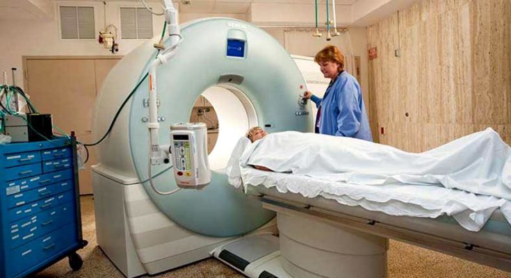
CT Scans – responsible for DNA damage? Image Courtesy – ttps://healthinformatics.wikispaces.com.
Utilizing new lab innovation, researchers have demonstrated that cell damage is perceptible after patients were undergone CT scan. A new research performed at the Stanford University School of Medicine, it was found that low-dose radiation for CT scanning of cardiac and vascular patients effects on human cellular structure.
One of the leading authors of the research Patricia Nguyen told that is was known that subjection to low amount of radiation from ‘computed tomography scanning’ was responsible for cell damage. Dr. Patricia Nguyen, who was also the Assistant Professor of Cardiovascular Medicine at Stanford, told that though it was not comprehensible that low dose radiation was directly related to cancer or any negative effect in a patient’s body, but doctors should cohere to dose reduction strategies.
The research has been published in the “Journal of the American College of Cardiology – Cardiovascular Imaging”. Other two leading writers of this research paper were Won Hee Lee, PhD and Yong Fuga Li, PhD.
Read: New technology for tooth rejuvenation will come very soon
Dr. Joseph Wu, MD and senior author of the research told that utilization of medical imaging for heart disease was at a vast range during the past 10 years. Dr. Joseph Wu, who was also a professor of ‘medicine and radiology’, and also the Director of the ‘Stanford Cardiovascular Institute’, also told that those ‘low-dose radiation’ although used to expose patients with not a significant amount, but, what was the actual effect on patient’s body due to those low-dose radiation was not known to anybody. Dr. Joseph Wu clarified that they have made the technology which could permit them to observe the cellular changes at very tenuous level.
Alongside the blossoming utilization of cutting edge medicinal imaging tests over the past 10 years have come in rising general health worries about conceivable connections between ‘low-dose radiation’ and malignancy. The anxiety is that expanded radiation exposure from such diagnostic strategies like CT scans, which open the body to low-dosage X-ray beams, can harm DNA and make transmutation that stimulus cells to develop into malignancy.
Dr. Joseph Wu told that presently there has been restricted logical proof which could demonstrate the impacts of this ‘low-dose radiation’ on human body, as indicated by the research. A bill has been focussed through Congress to provide more funds for research on health impacts of ‘low-dose radiation’. The current research indicated more research on the subject.
Read: Mosquito Bites may be a Gene factor
In this study, specialists analyzed the consequences for human cells of ‘low-dose radiation’ from an extensive variety of cardiovascular and vascular CT scans. These imaging methods are ordinarily utilized for various reasons, including administration of patients associated with having obstructive coronary artery sickness, and for those with ’aortic stenosis’, in arrangement of ‘transcatheter aortic valve’ substitution.
The research stated that a CT scan, which is utilized for imaging and diagnostic methods all through the body, uncover patients to no less than 150 times the measure of radiation in comparison to a solitary chest X-ray. The National Cancer Institute of United States evaluated in the year 2007 that 29,000 future malignancy cases could be allocated with the aftermath of 72 million CT scans performed in the United States that year.
Researchers inspected blood samples of 67 patients experiencing heart CT angiograms. Utilizing such systems as entire genome sequencing and ‘flow cytometery’ to gauge biomarkers of DNA harm; researchers analyzed blood samples of patients both previously, and after experiencing CT angiograms. The research demonstrated a hike in DNA damage and cell death, and also increased activities of genes necessitated in cell repair and death. The study also observed that most of the damaged cells were repaired, a little amount of cells died.
Read: Top 10 Medical achievements of the century
The research revealed that radiation exposure from ‘cardiovascular CT angiography’ might bring about DNA harm that could propel changes if damaged cells were not repaired or disposed of appropriately; and it has also been observed that progressive cell death after ‘repeated exposures’ might cause danger.
Dr. Patricia Nguyen told that they need to learn more because the effect was not amiable at these ‘low-dose radiation’, though the research supported the concept that doctors should not use the best quality images in all the cases. CT scans could not be eliminated because of its immense importance in diagnosis system, but doctors could make it safer by decreasing its doses. Doctors could utilize upgraded machines and technology to protect their patients.
A very important note has been added by Dr. Patricia Nguyen was that they did not notice any ‘DNA damage’ with patients of average weight and regular heart-bit under the situation of ‘lowest doses of radiation’.
Reference: Sciencedaily


